-
Home

-
Quick Links
-
Blog
- Read More
Nurse Practitioner Week: Ways to Celebrate the Advanced Practice Providers on Your Neuro Team
Nurse Practitioner Week will be held November 8-14, 2026, and gives your neuro unit a chance to recognize its advanced practice providers. Your neuro nurse practitioners handle complex cases every single day, often without much fanfare. National Nurse Practitioner Week gives you a clear reason to show your appreciation this year.
A handwritten note, a shared meal, or public recognition at a staff meeting can go a long way. The following ideas walk you through meaningful ways to honor the advanced practice providers on your neuro team.
How Can You Celebrate Nurse Practitioner Week on Your Neuro Team?
You can celebrate Nurse Practitioner Week with simple recognition, professional development support, and public appreciation. Start with a personal thank you, then add a team breakfast or a shout-out in your newsletter. Pair the gesture with support for a conference or certification to show lasting investment in their growth.
Nurse Practitioner Week Ideas for Everyday Recognition
Nurse Practitioner Week gives your unit an easy opportunity to show real appreciation. Your advanced practice providers juggle diagnostics, treatment plans, and patient education around the clock. A few thoughtful gestures throughout the week remind them their work gets noticed.
- Post a written thank-you note on each NP's workstation
- Bring in breakfast or coffee for the team on a set morning
- Share a shout-out during a staff huddle or in a department newsletter
- Ask physicians and other team members to submit short thank-you messages
Recognition works best when it highlights specific contributions instead of generic, catch-all praise. For instance, thank an NP by name for a fast diagnosis or a difficult family conversation handled well. A closer look at how nurse practitioners lead neuro-critical care teams shows why that daily impact deserves recognition.
Recognition should extend beyond one single week whenever your budget and schedule allow it. Consider setting a recurring quarterly reminder to recognize one NP's achievement or milestone. A regular habit like this turns one week of appreciation into part of your everyday team culture.
Peer nominations add another layer of meaning to any Nurse Practitioner Week celebration you plan. Invite nurses and support staff to nominate an NP whose work made a real difference this year.
Support Professional Growth Beyond the Celebration
Recognition matters, but real appreciation always goes further than a single celebration week. Your neuro nurse practitioners want opportunities to grow their clinical skills and their careers. Nurse Practitioner Week gives you a natural moment to talk about their next steps.
Ask each NP what certification or conference would help them the most this year. Then bring that request to your budget planning meeting with specific numbers attached.
Leadership development matters just as much as clinical training for advanced practice providers. Nurses who step into mentorship or committee roles build skills that benefit the whole unit. AANN's piece on building a neuroscience center of excellence through APP leadership offers a helpful starting point for that conversation.
Career growth conversations also boost retention among your advanced practice staff over time. Nurse practitioners who feel invested in tend to stay longer and mentor others in turn. Stability like this strengthens patient care across your entire neuroscience unit long term.
Make It a Launchpad for Ongoing Support
Nurse Practitioner Week works best as a starting point, not a finish line. Use the week to gather feedback on what your NPs actually need to succeed. A short survey or a casual conversation can surface ideas you had not considered.
Some NPs want more scheduling flexibility, while others want funding for a certification exam. Write down every suggestion, so your team sees the celebration turn into real action.
Follow through on at least one request within the following month if you can. Small, visible wins build trust faster than a long list of unmet promises. For extra ideas, AANN's guide on investing in excellence through nursing professional development covers events and strategies worth exploring.
Budget cycles vary, so timing your request around Nurse Practitioner Week can help it land faster. Managers often approve smaller asks tied to a specific, well-timed occasion like this one.
A well-supported NP delivers better care and stays with your organization for years. Nurse Practitioner Week gives you the perfect reason to start that conversation today. Treat it as the beginning of a year-round commitment to your advanced practice team.
Start Planning Your Nurse Practitioner Week Celebration Today
Nurse Practitioner Week gives your neuro unit a simple, meaningful reason to pause and say thank you. Small gestures build trust, and bigger investments in growth build loyalty that lasts. Your advanced practice providers deserve both kinds of recognition throughout the entire year.
Every neuroscience nurse practitioner on your team shapes patient outcomes in ways that often go unseen. AANN offers education, certification pathways, certificate programs, and community support to help them keep growing. Reach out to AANN to explore resources that support the neuroscience NPs on your team.
Categories: Professional Development
Making the Case to Your Manager for Conference Funding
You gain skills at neuroscience conferences that no other training can match, but funding is not automatic. Your manager controls a limited education budget and needs a clear reason to approve it. A strong, well-prepared pitch turns a hesitant maybe into a fully approved request.
Managers respond well to numbers, clear outcomes, and a direct connection to patient care. Vague, unclear requests get pushed to the bottom of the pile every single time. The following approach gives you a practical way to ask for conference funding and get a yes.
Why Neuroscience Conferences Matter for Your Career and Your Unit
Neuroscience conferences give you access to research and techniques not covered in daily practice. Sessions on stroke care, epilepsy management, and spine health sharpen skills you use every shift. Networking with other neuroscience nurses also opens doors to mentorship and career growth.
Your unit benefits directly when you bring new knowledge back to the floor. A nurse who attends a conference often becomes an informal trainer for the rest of the team. Knowledge like this spreads quickly and raises the skill level of your entire team.
Professional development also reduces burnout and improves long-term retention across every neuroscience unit. Nurses who feel supported in their growth tend to stay in their roles longer. AANN's guide to investing in excellence through nursing professional development explains why ongoing education pays off for entire units.
Conference attendance also builds your professional credibility. Many neuroscience conferences offer contact hours that count directly toward CNRN or SCRN recertification.
Build a Budget Request Your Manager Cannot Ignore
A strong funding request starts with specific numbers, not general enthusiasm. List registration fees, travel costs, lodging, and time away from the unit up front. Managers trust requests that show you have already done the homework.
Next, connect the conference directly to a unit goal or a known gap in care. For example, tie a stroke session to your unit's current door-to-needle time initiative.
Include a plan for sharing what you learn when you return to work. Offer to present a short summary at a staff meeting or write a one-page recap. AANN's piece on strategies for success as a new stroke coordinator shows how shared learning strengthens an entire unit.
Attach a list of neuroscience conferences with dates, costs, and relevant sessions highlighted. A clear comparison like this makes the decision easier for a manager juggling multiple requests. Ask for a specific dollar amount instead of leaving the number open to negotiation.
Time Your Neuroscience Conferences Request Around Key Dates
Timing changes how likely your manager is to approve neuroscience conferences funding for the year. Most facilities set education budgets at the start of a new fiscal year cycle. You submit your funding request as soon as the new budget period opens.
|
Key Date |
What It Means |
|
Start of new fiscal year |
Your facility opens fresh education budgets for conference funding requests |
|
November |
AANN Annual Conference registration usually opens, often with lower early pricing |
|
March 13-16, 2027 |
The AANN Annual Conference takes place in Seattle, themed Uniting Expertise, Elevating Care |
You can reference AANN's Annual Conference page for updated pricing, sessions, and speaker details. If your manager hesitates, offer a smaller ask first, such as a single day pass. You follow up after the event with a report, similar to AANN's stroke certification article. Need help convincing your boss you should attend? Use our customizable approval letter to request funds to attend or time off.
Make Your Case for Neuroscience Conferences Today
Neuroscience conferences pay off for you, your unit, and your patients when you make a clear case. Managers say yes more often when they see specific costs, goals, and a plan to share what you learn. Preparation turns your request from a hopeful ask into an easy decision.
AANN's Annual Conference and other neuroscience events offer education you cannot find anywhere else. Your growth as a nurse strengthens the entire neuroscience unit around you every day. Contact your manager this week to start the conversation about funding your next conference.
Categories: Professional Development | Education | Leadership
How Do You Turn a Unit Problem Into a Research Study?
A small frustration during an ordinary shift often becomes the seed for nursing research topics. A workaround you use every day may point to a real gap in current practice. Turning that observation into a formal study takes structure, but the process is more approachable than it seems.
Every unit has real problems worth studying, from documentation gaps to inconsistent handoffs. The right question, a review of existing evidence, and some mentorship can carry an idea forward. The sections below walk through the steps for turning a unit problem into a research study.
How Do You Turn a Unit Problem Into a Nursing Research Topic?
You turn a unit problem into a research study by defining the issue clearly and checking existing evidence first. Frame the problem as a specific, answerable question instead of a general frustration. From there, a simple design and a mentor's guidance can carry your idea into a real nursing research topic worth pursuing.
Spot Nursing Research Topics Hiding in Everyday Problems
How do you determine which topics to even look further into when nothing seems immediately apparent? Uneven documentation practices between nurses can often signal a genuine research opportunity worth exploring. Ask your colleagues whether they notice the same issue in their own daily work. A shared frustration across the unit usually points to a stronger research question.
Before you commit to a topic, search for existing literature on the same problem. Someone else may have already studied it, which can save you significant time. AANN's article on standardizing neurocheck documentation for improved outcomes shows how one unit-level problem became a published nursing research topic.
If the literature is thin or conflicting, your problem may be worth new research. Gaps in the evidence often make the strongest and most publishable research questions.
Turn Your Idea Into a Clear, Answerable Research Question
A strong research question stays specific, measurable, and realistic for your setting. Broad questions like improving patient safety rarely lead anywhere useful on their own. Narrow the focus to one population, one intervention, and one measurable outcome.
The PICO format, covering patient, intervention, comparison, and outcome, helps sharpen a vague idea. Writing your question in this format also clarifies what data you will need to collect.
Talk with a nurse researcher or an academic partner early in the overall process. Their feedback can catch design problems before you invest significant time in data collection. AANN's piece on building a neuroscience center of excellence through APP leadership highlights how mentorship strengthens research work.
Small, focused pilot studies work well for a first attempt at nursing research. A short, well-designed pilot can demonstrate value before you pursue a larger study. Many productive research careers begin with a single unit-level project like this one.
Get Support to Pursue Further
Pursuing nursing research topics gets easier once you know where to find support. Most hospitals have a research or evidence-based practice council you can approach for guidance. Academic partnerships with local nursing schools also offer mentorship and statistical help.
Professional organizations also provide another valuable layer of support for new researchers just starting out. AANN, for example, connects nurses with resources built specifically for neuroscience nursing research.
The Virtual International Neuroscience Nursing Research Symposium gives you a platform to present early findings and get feedback. Submitting a poster or abstract there can turn a unit-level project into recognized nursing research.
Funding does not need to be a major barrier to getting started either. Many facilities and professional organizations offer small grants for nurse-led research projects each year.
Documentation and neuro assessment projects translate especially well into strong nursing research topics. AANN's guide on neuro assessment, from brain anatomy to seizure management, shows how those skills connect to research design. Your everyday clinical judgment already gives you a strong foundation for meaningful research.
Turn Your Next Unit Problem Into Nursing Research
Every unit problem you notice carries the seed of a meaningful nursing research topic. A clear question, a review of existing evidence, and the right mentor can carry that seed forward. You do not need a doctorate or a large budget to start contributing real evidence.
AANN supports nurses at every stage of the research process, from a first idea to a symposium presentation. Your bedside experience gives you insight that many researchers never have access to. Get in touch with AANN to find the resources and community behind your next nursing research topic.
Categories: Professional Development | Leadership
Common Myths About Concussions and What the Evidence Shows
Patients, families, and even clinicians build their view of head injury care around concussion myths. Many of these ideas sound reasonable, yet they collapse under close scientific review. You can guide patients toward a safer recovery once you know what the evidence shows.
Outdated advice about imaging, rest, and long-term brain damage still circulates widely today. Protect patients from unnecessary fear and delayed treatment by correcting these myths. The list below walks through five common concussion myths and the research that contradicts them.
What Does the Evidence Show About Common Concussion Myths?
The evidence shows that most concussion myths do not survive close scientific examination. Concussions do not always involve a loss of consciousness, and imaging often looks normal. Recovery timelines also vary widely, so no single myth fits every patient's experience.
Concussion Myth 1: A Blackout Always Confirms a Concussion
Concussion myths spread quickly because head injuries often seem simple on the surface. Many patients and families believe a concussion always causes a blackout or visible wound. Neither assumption holds up once you look at the actual research on head injuries. 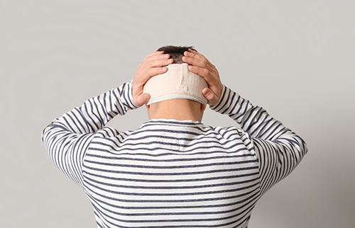
Most concussions involve no loss of consciousness, and visible injury is not required. You may still watch closely for symptoms like confusion, headache, or sensitivity to light. Assess every head injury carefully, regardless of whether the patient lost consciousness. You risk delaying diagnosis and proper care by waiting for a dramatic symptom. A thorough evaluation catches concussions that a quick glance would otherwise miss entirely.
Concussion Myth 2: A Normal CT Scan Rules Out an Injury
Another common myth claims that a normal CT scan rules out a concussion completely. Concussion is a functional injury, so standard imaging often looks normal despite real symptoms. You cannot rule out a concussion using imaging results alone in most clinical cases.
You risk leaving a genuine concussion undiagnosed for several weeks by trusting imaging alone. A thorough clinical exam catches symptoms that a scan on its own would miss. Combining imaging with a full symptom history and a hands-on neurological exam is the best option.
Patients deserve a full evaluation even when their scan results come back clear. Thorough documentation of the exam helps disprove common concussion myths about imaging accuracy.
Concussion Myth 3: Complete Rest Speeds Up Recovery
A third myth suggests that complete rest, with no activity, speeds recovery fastest. Older guidance recommended strict rest in a dark room for several days straight. Newer research shows this old approach can slow recovery for many patients today.
Current guidance favors a gradual return to light activity within a few days. Most patients benefit from short walks within twenty-four to forty-eight hours. Screen time and reading in moderation no longer carry the strict bans they once did. AANN's overview of standardizing neurocheck documentation for improved outcomes shows how careful assessment supports better recovery.
Concussion Myth 4: One Injury Causes Permanent Brain Damage
Some patients believe a single concussion causes permanent brain damage on its own. Most people recover fully within weeks when they follow a structured recovery plan. Repeated concussions, however, do raise the risk of longer term cognitive problems later.
Track the number and timing of prior concussions for every returning patient. Spacing between injuries matters more for long term outcomes than the injury count alone. AANN's guide on neuro assessment, from brain anatomy to seizure management, offers a useful framework for structured evaluation. Structured evaluation helps you catch subtle cognitive changes before they turn into lasting problems.
Concussion Myth 5: Symptoms Mimic a Stroke or Seizure
Some patients assume concussion symptoms look nothing like a stroke or a seizure. Symptom overlap between these conditions fuels real confusion for patients and their families. Other patients mistake concussion symptoms for a stroke or a seizure, which raises anxiety. atch closely for one-sided weakness, slurred speech, or sudden vision loss. Signs like these point toward a stroke rather than a typical concussion case. AANN's article on educating patients about the warning signs of a mini stroke offers a useful comparison.

How You Correct Concussion Myths in Practice
Correcting concussion myths works best through an open conversation, not a one-sided lecture. You ask patients and families what they already believe about typical recovery timelines. Their answers reveal exactly which myths you need to address first with them.
Use plain, everyday language instead of clinical terms when explaining the evidence. Written take-home materials reinforce what you say and reduce confusion later at home.
Aligned messaging across your entire care team prevents mixed signals for every patient. AANN's resource on silent strokes and future infarct risk shows how myths about one condition delay care for another.
Share the Evidence on Concussion Myths With Your Team
Concussion myths persist because outdated advice still circulates among patients and their clinicians. The evidence tells a different story about imaging, rest, and long-term recovery. Sharing accurate, evidence-based information protects your patients from unnecessary fear and delayed treatment.
Your role as a neuroscience nurse puts you in a strong position to correct these myths. Clear, evidence-based education builds trust and improves outcomes for every patient you see. Reach out to your neuroscience nursing network to keep these conversations going strong.
Categories: Education | Neurotrauma
Building a Neuroscience Center of Excellence Through APP Leadership
Hospitals nationwide depend on strong clinical leaders to build a true center of excellence. Advanced practice providers bring the clinical depth and vision that neuroleadership demands today. Their strategic mindset shapes how neuroscience programs grow stronger and more resilient over time. Building this kind of center takes more than skilled clinicians working in separate corners of a unit. It requires a shared vision, clear communication, and leaders who understand both bedside care and strategy.
Defining Neuro-Leadership Within Neuroscience Programs
Neuro-leadership blends clinical judgment with the strategic skills needed to guide entire departments forward. Advanced practice providers often sit between bedside nurses and hospital administration, translating clinical needs into policy. Because of this position, they shape protocols, staffing models, and quality improvement projects across neuroscience units.
Additionally, neuro-leadership requires ongoing education that goes beyond a provider's original degree program. Many APPs pursue the Neuro-APP Certificate Program to sharpen these skills. The certificate program covers clinical management alongside leadership development topics tailored to neuroscience practice settings.

Strong neuro-leadership also depends on mentorship between experienced APPs and newer team members. As a result, mentorship spreads institutional knowledge and builds a pipeline of future clinical leaders.
Programs that invest in mentorship also see measurable gains in staff retention over time. Newer providers gain confidence faster when paired with an experienced neuroleadership mentor. Therefore, neuroscience departments should treat mentorship as a core part of their leadership strategy.
How APP Leadership Strengthens Clinical Outcomes
Advanced practice providers coordinate care across specialties, which reduces delays in treatment for neurological patients. For example, an APP leading a stroke coordinator role can shorten the time between diagnosis and intervention. Faster coordination often means better recovery outcomes for patients facing time-sensitive neurological conditions.
Furthermore, APPs standardize protocols that steady patient care when multiple providers rotate through a unit. Standardized protocols reduce variation in treatment approaches, which supports safer and more predictable patient outcomes overall.
APP-led programs also track outcomes closely, and neuro-leadership drives this ongoing discipline across teams.
- Reviewing readmission rates and identifying patterns tied to specific care gaps
- Auditing documentation to confirm neurological assessments happen on schedule
- Adjusting staffing models based on real patient acuity data
- Sharing outcome trends with the wider care team during regular meetings
Such structured approaches turn raw data into practical changes that improve patient care daily. Meanwhile, teams gain confidence knowing decisions rest on evidence rather than guesswork alone. Therefore, this kind of ongoing professional development makes APP leadership measurable rather than anecdotal within any neuroscience program.
Hospitals that adopt this approach often see fewer preventable complications across their neuroscience units, a trend explored in the Journal of Neuroscience Nursing. As a
result, patients benefit directly from the structure that strong APP leadership provides.
Building a Path Toward Neuro-Leadership
Nurses interested in neuro-leadership often start by pursuing an advanced practice degree and specialized clinical training. From there, many pursue the CNRN certification through the American Board of Neuroscience Nursing (ABNN) to demonstrate expertise. The CNRN credential represents specialized experience and knowledge in the care of patients with neurological trauma and illness.
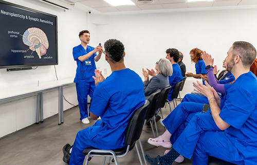
Professional development does not stop after earning a credential or finishing a degree program. Attending the AANN Neuroscience Nursing Annual Conference gives APPs fresh insight into leadership strategies used nationwide, and the conference has a track of sessions for APPs with advanced content.
Mentorship structures also strengthen this path, pairing early-career APPs with proven neuro-leadership role models. Mentor relationships often continue well past initial training programs into ongoing professional networks. Consequently, new leaders emerge with both technical skill and confidence in strategic decision-making.
Local chapters and special focus groups also provide additional support for nurses on this path. Getting involved early builds both skill and visibility within the broader neuroscience nursing community.
Growing Across Your Organization
Every neuroscience program can strengthen its leadership pipeline by investing in advanced practice providers early. Organizations that support ongoing education and mentorship see stronger neuro-leadership emerge from internal teams. Instead of searching externally for new hires, hospitals can grow the leaders they already employ.
Consider offering protected time for APPs to pursue certificate programs and attend conferences annually. Such investment pays off through improved retention, sharper clinical outcomes, and a stronger center of excellence overall.
Take the Next Step Toward Neuro-Leadership
Building a neuroscience center of excellence starts with recognizing the leaders already working within your organization. Strong neuro-leadership does not happen by accident, and it requires deliberate investment in people and process. Advanced practice providers who receive support today become the clinical leaders your program depends on tomorrow.
AANN remains committed to supporting neuroscience nurses at every stage of theirAANN remains committed to supporting neuroscience nurses at every stage of theirleadership journey. From certificate programs to conference education, resources existto help your team grow stronger together. We encourage program leaders andadvanced practice providers alike to reach out to AANN and start building that future today.
Categories: Professional Development | Advanced Practice | Neuro-APP Certificate
Recent Advances in Ischemic and Hemorrhagic Stroke Treatment
Faster recognition, better imaging, and refined interventions now shape how care teams respond to stroke. Over the past decade, stroke treatment has advanced considerably and improved outcomes for many patients. Neuroscience nurses sit atthe center of these advances, applying new tools at the bedside daily.
Ischemic and hemorrhagic strokes require very different treatment approaches despite sharing some early warning signs. Understanding these differences helps nurses support faster, safer care during a true medical emergency.
Treatment advances now include expanded time windows for clot-removing procedures and refined clot-dissolving medications. For ischemic stroke, mechanical thrombectomy has extended treatment options well beyond the traditional early hours. For hemorrhagic stroke, faster blood pressure management now helps limit additional bleeding and brain damage.
Advances in Ischemic Stroke Treatment
Ischemic stroke occurs when a clot blocks blood flow to part of the brain suddenly.Tissue plasminogen activator, or tPA, remains a cornerstone stroke treatment for eligible patients within set time windows. Recent research has extended eligibility forthis medication beyond the original three-hour treatment window.
Mechanical thrombectomy has also transformed ischemic stroke treatment for patients with large vessel blockages. Such a procedure physically removes the clot, often restoring blood flow within a critical treatment window. Because outcomes depend heavily on timing, rapid recognition remains essential for every care team member.
Additionally, some centers now use extended imaging protocols to identify patients who qualify for thrombectomy later. Such an approach helps more patients access treatment even outside traditional early time windows.
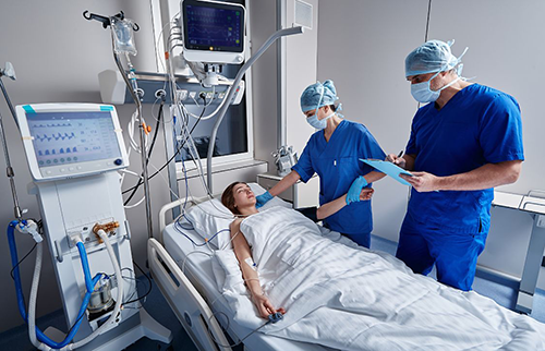
Nurses who earn the SCRN certification, offered by the American Board of Neuroscience Nursing (ABNN), understand these protocols closely. As a result, they help teams move quickly when every minute of stroke treatment counts.
Advances in Hemorrhagic Stroke Treatment
Hemorrhagic stroke happens when a blood vessel ruptures, causing bleeding directly into or around the brain. Because bleeding can worsen quickly, stroke treatment here focuses heavily on controlling blood pressure and swelling. Surgical intervention sometimes becomes necessary to relieve pressure and remove accumulated blood.
Furthermore, newer medications now help reverse blood thinners faster than older treatment protocols allowed. Such speed matters greatly for patients taking anticoagulants who experience a hemorrhagic stroke unexpectedly.
Consider the priorities that shape hemorrhagic stroke treatment during the first critical hours:
- Monitoring blood pressure closely and adjusting medications to reduce further bleeding risk
- Watching for signs of rising intracranial pressure throughout ongoing assessments
- Coordinating quickly with neurosurgery when surgical intervention becomes necessary
- Reversing anticoagulant effects promptly when patients arrive on blood thinners
Rapid, coordinated action during this window often determines a patient's long-term recovery potential. Therefore, neuroscience nurses must stay current on evolving protocols and medication options.

Ongoing research published through the Journal of Neuroscience Nursing tracks these developments closely. Reviewing this research regularly helps nurses apply the newest evidence at the bedside confidently.
Why Nursing Expertise Shapes Modern Treatment
Advances in stroke treatment only help patients when clinical teams recognize symptoms and respond quickly. Nurses often serve as the first point of contact when a patient shows signs of stroke. Because early recognition drives every subsequent treatment decision, nursing assessment skills remain critically important.
Consequently, hospitals increasingly invest in ongoing stroke education for their neuroscience nursing staff. Such investment helps teams apply new protocols correctly across every single shift.
Standardized neurological assessments also help teams track patient changes throughout the entire treatment process. As a result, care teams catch complications earlier and adjust treatment plans before problems escalate.
Stay Current on Stroke Treatment With AANN
Stroke treatment continues advancing quickly, and neuroscience nurses play a central role in applying that progress. From extended thrombectomy windows to faster hemorrhagic stroke interventions, the pace of change remains significant. Staying current with these advances helps nurses protect patient outcomes during every stroke emergency.Stroke treatment continues advancing quickly, and neuroscience nurses play a central role in applying that progress. From extended thrombectomy windows to faster hemorrhagic stroke interventions, the pace of change remains significant. Staying current with these advances helps nurses protect patient outcomes during every stroke emergency.
AANN offers education and resources built specifically for stroke care professionals. Such resources help nurses translate new research into confident, timely bedside decisions.
Categories: Stroke | Education
Differential Diagnosis of Obstructive vs Normal Pressure Hydrocephalus
Cerebrospinal fluid, or CSF, can build up within the brain's ventricles and lead to hydrocephalus over time. This buildup raises pressure inside the skull and can damage brain tissue if left untreated. Nurses play a central role in recognizing the signs that point toward this serious condition.
Not all hydrocephalus looks the same, and the underlying cause changes how nurses should respond. Obstructive and normal pressure hydrocephalus, or NPH, present differently despite some overlapping symptoms. Readers will find a clear walk-through of the clinical clues that separate these two forms.
How Obstructive and Normal Pressure Differ
Obstructive hydrocephalus results from a physical blockage that stops cerebrospinal fluid from draining normally. Normal pressure hydrocephalus instead involves impaired fluid absorption despite pressure readings that often appear normal. Because the causes differ, nurses must rely on distinct clinical signs to tell these forms apart.
Understanding Hydrocephalus and Its Two Primary Forms
Hydrocephalus develops when cerebrospinal fluid production outpaces the body's ability to drain it properly. In obstructive cases, a tumor, cyst, or structural abnormality blocks normal fluid flow through the ventricles. Such blockage often causes pressure to rise quickly, which creates a more urgent clinical picture.
Normal pressure hydrocephalus, however, tends to develop gradually and often affects older adults specifically. Fluid absorption slows over time, yet intracranial pressure readings frequently remain within a normal range. Because symptoms develop slowly, families and clinicians sometimes mistake early signs for normal aging changes.

Additionally, obstructive hydrocephalus can appear at any age, including in infants born with structural abnormalities. NPH almost always appears in adults over the age of sixty, which helps narrow the diagnosis.
Understanding these differences helps nurses set appropriate expectations for treatment and recovery timelines. Obstructive cases often require urgent surgical intervention, while NPH management can follow a more measured pace.
Key Clinical Signs That Point to Each Diagnosis
Obstructive hydrocephalus often presents with a classic triad of symptoms that develop over a short period. Patients typically report severe headaches, nausea, and vomiting alongside rapidly worsening vision or consciousness changes. Because these symptoms escalate quickly, it requires immediate clinical attention and imaging.
Normal pressure hydrocephalus instead presents with a different, slower triad that nurses should learn to recognize. Nurses recognize this pattern through gait disturbances, cognitive decline, and urinary incontinence developing over months or years.
Consider how nurses distinguish these presentations during a thorough clinical assessment:
- Noting whether symptoms developed suddenly or gradually over an extended period
- Assessing gait changes, since NPH often causes a distinct shuffling walk pattern
- Reviewing cognitive changes alongside any reported memory or attention difficulties
- Checking for incontinence patterns that may point toward normal pressure hydrocephalus
Careful documentation of these patterns helps physicians choose the right diagnostic path forward. Furthermore, accurate early observations from nursing staff often shape how quickly treatment begins.
Nurses who sharpen these assessment skills through continued education support faster, more accurate diagnoses. Therefore, ongoing clinical training remains essential for anyone working with neurological patients regularly.
Diagnostic Tools Nurses Use to Confirm Hydrocephalus
Computed tomography, or CT, scans often serve as the first imaging step when hydrocephalus is suspected. Such scans reveal enlarged ventricles quickly, which helps confirm whether fluid buildup has occurred. Magnetic resonance imaging, or MRI,then provides more detailed information about the underlying cause.

For suspected NPH cases, physicians may also order a lumbar puncture to test symptom response. Improvement after fluid removal often supports an NPH diagnosis and helps guide treatment decisions.
Supporting Accurate Hydrocephalus Diagnosis Through Nursing Education
Nurses who understand hydrocephalus deeply contribute meaningfully to faster, more accurate diagnostic pathways. The Journal of Neuroscience Nursing regularly publishes research on emerging diagnostic approaches for this condition. Staying current with this research strengthens clinical judgment across every neuroscience care setting.
Pursuing the CNRN certification through the American Board of Neuroscience Nursing (ABNN) also builds this expertise. Many nurses further sharpen these skills through the AANN Neuroscience Nursing Annual Conference each year.
Strengthen Your Hydrocephalus Assessment Skills With AANN
Recognizing the difference between obstructive and normal pressure hydrocephalus takes sharp clinical observation and ongoing training. Nurses who master these distinctions help patients receive faster, more accurate treatment when it matters most. Such clinical skill remains one of the most valuable tools in neuroscience nursing practice.
AANN provides education, certification pathways, and community resources built to strengthen this exact type of expertise. Review the Neuroscience Resources page of the AANN website for quick guides and toolkits.
Categories: Hydrocephalus | Professional Development | Education
How Nurse Practitioners Lead the Neuro-Critical Care Team
A nurse practitioner, or NP, often serves as the steady hand guiding a neuro-critical care team. Patients in these units face rapid changes that demand fast, informed clinical decisions around the clock. Nurse practitioners bring the training and presence needed to manage that constant pressure well.
Neuro-critical care differs from general critical care because brain and spinal cord injuries shift quickly. Small changes in a patient's condition can signal serious complications if no one catches them early.
What a Nurse Practitioner Does on a Neuro-Critical Care Team
A nurse practitioner monitors neurological status, adjusts treatment plans, and communicates changes across the care team. On a neuro-critical care unit, this often means frequent neuro-checks and rapid documentation of subtle shifts. Because conditions change quickly, the nurse practitioner becomes the steady link between physicians, nurses, and families.
The Nurse Practitioner Role in Neuro-Critical Care
Nurse practitioners working in neuro-critical care manage patients recovering from stroke, brain injury, and spinal trauma. Their training allows them to interpret imaging results, adjust medications, and order additional testing quickly.
Nurse practitioners also lead family conversations during some of the most difficult moments in care. Families often look to the nurse practitioner for clear updates on prognosis and next steps. Clear communication reduces confusion and builds trust between families and the wider clinical team.
Additionally, nurse practitioners often supervise newer staff working within the neuro-critical care unit. Mentorship helps junior nurses understand subtle neurological changes before they become serious problems.
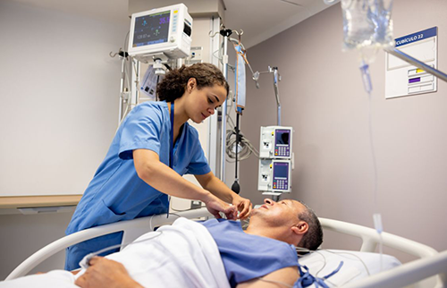
Many nurse practitioners in this specialty pursue the CNRN certification through the American Board of Neuroscience Nursing (ABNN). The CNRN credential represents specialized experience and knowledge in the care of patients with neurological trauma and illness. As a result, patients and colleagues gain added confidence in that provider's clinical judgment.
Daily Responsibilities Inside the Neuro-Critical Care Team
Every shift brings a fresh set of decisions that shape patient outcomes across the unit. A nurse practitioner typically starts the day reviewing overnight neurochecks and any new imaging results. From there, priorities shift quickly based on which patients show signs of neurological change.
Furthermore, nurse practitioners coordinate closely with therapists, pharmacists, and case managers throughout each shift. Close coordination keeps recovery plans on track even as a patient's condition evolves.
Consider the range of tasks a nurse practitioner manages during a single twelve-hour shift:
- Performing detailed neurological exams at set intervals throughout the day
- Adjusting sedation and pain management plans based on patient response
- Reviewing lab values and imaging for signs of new complications
- Updating families and physicians on any meaningful clinical changes
These responsibilities require sharp clinical judgment paired with calm, clear communication under pressure. Because the stakes remain high, nurse practitioners rely on strong training and ongoing education to stay sharp.
Continuing education through resources like the Journal of Neuroscience Nursing keeps clinical knowledge current. Staying current with new research helps nurse practitioners adjust their practice as evidence evolves.
Why Nurse Practitioner Leadership Improves Patient Outcomes
Research on advanced practice providers shows that nurse practitioner involvement often improves recovery timelines significantly. When a nurse practitioner leads daily rounds, care teams tend to catch complications earlier than before. Earlier detection frequently means faster intervention and better outcomes for patients with neurological injuries.
Therefore, hospitals increasingly place nurse practitioners at the center of neuro-critical care staffing models. Such change reflects growing recognition of the clinical value these providers bring to complex units.

Nurse practitioners also reduce strain on physician staffing by handling routine assessments and adjustments independently. Consequently, physicians can focus their attention on the most complex or unstable patients in the unit.
Building Stronger Nurse Practitioner Teams Over Time
Hospitals looking to strengthen their neuro-critical care staffing should invest in nurse practitioner training and mentorship. Supporting attendance at the AANN Neuroscience Nursing Annual Conference helps providers stay current with best practices as conference sessions increasingly address neuro-critical care leadership directly.
Encouraging membership through AANN also connects nurse practitioners with a wider professional community. Such connection supports long-term growth and keeps clinical practice aligned with evolving standards of care.
Partner With AANN to Strengthen Your Neuro-Critical Care Team
Nurse practitioners bring clinical depth, steady leadership, and constant presence to neuro-critical care units nationwide. Their role continues to grow as hospitals recognize the value of advanced practice expertise at the bedside. Supporting these providers with education and mentorship strengthens outcomes for every patient in their care.
AANN offers conference education, certification pathways, and community connections built specifically for this specialty. From the Annual Conference to ongoing professional resources, support exists for nurse practitioners at every career stage. If your team wants to strengthen neuro-critical care leadership, contact AANN to explore these resources together.
Categories: Critical Care | Advanced Practice | Professional Development | Leadership
Emergency Management of Autonomic Dysreflexia in Spinal Cord Injury
Autonomic dysreflexia is a medical emergency. It affects people with spinal cord injuries at the T6 level or higher. Without fast action, it can cause a stroke, seizure, or even death. Every nurse who cares for spinal cord injury patients must recognize it immediately.
This condition happens when something painful or irritating occurs below the level of the spinal cord injury. The body reacts with a sudden and dangerous spike in blood pressure. Because the injury blocks normal nerve signals, the body cannot control the response.
The good news is that autonomic dysreflexia is treatable. With quick thinking and the right steps, nurses can resolve an episode and prevent serious harm. The American Association of Neuroscience Nurses (AANN) provides clinical resources to help nurses manage conditions like this with confidence.
Who Is at Risk for Autonomic Dysreflexia?
Not every spinal cord injury patient is at risk. Autonomic dysreflexia specifically occurs in patients with injuries at or above the T6 spinal level. That includes most cervical (neck) and upper thoracic (upper back) injuries.
The higher the level of injury, the greater the risk. For example, a patient with a C4 injury is at higher risk than a patient with a T5 injury. However, both groups need close monitoring.
Interestingly, patients with incomplete spinal cord injuries can also develop autonomic dysreflexia. Therefore, nurses should never assume a patient is safe from this condition simply because their injury is incomplete.
Recognizing the Signs of Autonomic Dysreflexia
Speed is everything. Knowing the symptoms allows nurses to act before the blood pressure reaches a dangerous level. Common signs include:
- Sudden high blood pressure – Systolic BP 20 to 40 mmHg above the patient's normal baseline
- Pounding headache – Often severe and sudden
- Sweating above the injury level – Especially on the face and neck
- Flushing or blotchy skin above the injury level
- Slow heart rate (bradycardia) or fast heart rate
- Stuffy nose
- Goosebumps below the injury level
- Anxiety or a feeling that something is very wrong
It is important to know each patient's baseline blood pressure. Many spinal cord injury patients have naturally low blood pressure. A reading of 110/70 mmHg could actually be dangerously high for them. Always compare to the individual baseline, not general normal values.
What Triggers Autonomic Dysreflexia?
The most common trigger is a full or blocked bladder. In fact, bladder problems cause up to 85% of all autonomic dysreflexia episodes. However, any painful or uncomfortable stimulus below the injury level can trigger it.

Other common triggers include:
- A blocked or kinked urinary catheter
- Constipation or a full bowel
- A pressure injury or skin breakdown
- Tight clothing, shoes, or compression devices
- A urinary tract infection
- An ingrown toenail or undetected fracture
- Menstrual cramps or labor in female patients
Because patients may not feel pain below the injury, they often do not know what is causing the episode. The nurse must act as their detective and systematically check for each possible trigger.
Step-by-Step Emergency Management of Autonomic Dysreflexia
When autonomic dysreflexia is suspected, act immediately. Follow these steps in order:
Step 1: Sit the Patient Upright
First, raise the head of the bed as high as it will go. This helps lower blood pressure naturally through gravity. Also remove any tight clothing, binders, or compression stockings right away.
Step 2: Check Blood Pressure
Next, take the blood pressure immediately. Monitor it every two to five minutes. Call the doctor or advanced practice provider right away. Do not wait to see if it comes down on its own.
Step 3: Check the Bladder First
The bladder is the most likely cause. If the patient has a urinary catheter, check for
kinks, blockages, or displacement. Straighten any kinks and ensure the bag is draining freely. If the patient does not have a catheter, place one now. Use anesthetic lubricant
to reduce additional stimulation.
Step 4: Check the Bowel
If the bladder is not the cause, check for bowel problems. Use anesthetic ointment before any bowel assessment or manual removal of stool. This is important because
touching the area without anesthetic can make the blood pressure spike even higher.
Step 5: Look for Other Triggers
After that, systematically check for other triggers. Look at the skin from head to toe
below the injury. Check clothing, positioning devices, and any equipment touching the patient. Even a small wrinkle in a sheet can cause an episode.
Step 6: Give Medication If Needed
If the trigger is removed but blood pressure stays dangerously high, medication may
be needed. Common options include nitroglycerin paste or a fast-acting calcium channel blocker. Always follow the doctor's orders and your hospital's protocol. Document the drug, dose, time, and the patient's response.
Step 7: Monitor and Document
Finally, keep watching the patient for at least 30 to 60 minutes after the episode resolves. Document everything: the suspected trigger, each step taken, the timeline, and how the patient responded. This documentation helps prevent future episodes.
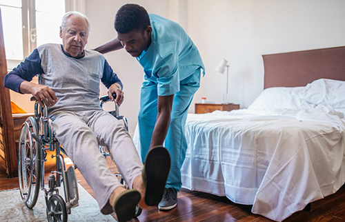
Preventing Autonomic Dysreflexia Before It Starts
Prevention is always better than treatment. Nurses play a key role in stopping autonomic dysreflexia from happening in the first place. A consistent bowel and bladder routine is the most effective preventive strategy.
Patient and family education is equally important. Patients who go home with a spinal cord injury need to know the signs of autonomic dysreflexia. They also need to know what to do before the ambulance arrives. AANN supports nurses in delivering this kind of education through its membership resources and continuing education programs.
Advance Your Skills With AANN's Spine Certificate Program
For nurses who care for spinal cord injury patients regularly, specialized training makes a real difference. The Spine Certificate Program offers structured, evidencebased learning.
Additionally, the AANN blog regularly publishes clinical content on spinal cord injury care and other neuroscience topics. Staying informed is one of the most powerful tools in a nurse's toolkit.
Categories: Spine | Professional Development
The Advanced Practice Impact: How the Neuroscience Nurse Practitioner Transforms Patient Outcomes
Every November, National NP Week celebrates the advanced practice nurses whoEvery November, National NP Week celebrates the advanced practice nurses whochange lives. Among them, the neuroscience nurse practitioner stands out. These professionals care for some of the most complex patients in healthcare. Their impact on patient outcomes is real, measurable, and significant.
Neurological diseases are growing in number every year. Yet there are not enough neurologists to meet the demand. That is where neuroscience nurse practitioners step in. They help close the gap between patients who need specialty care and the specialists available to provide it.
The American Association of Neuroscience Nurses (AANN) supports these advancedpractice nurses. AANN offers several educational resources for advanced practice providers, including content at the Neuroscience Nursing Annual Conference, the Neuro-APP Certificate Program, and Quick Guides.
What Does a Neuroscience Nurse Practitioner Do?
A neuroscience nurse practitioner is more than just a regular nurse. They haveadvanced clinical training. They are qualified to diagnose, treat, and manageneurological conditions. Their work covers a wide range of responsibilities.
For example, a neuroscience NP may:
- Perform full neurological exams to track a patient's brain and nerve function
- Order and review brain scans like MRIs or CT scans
- Manage medications for conditions such as epilepsy, stroke, and Parkinson's disease
- Coordinate care with surgeons, therapists, and social workers
- Educate patients and families on managing their condition at home
In short, neuroscience NPs bridge clinical care and patient support. They are a trusted constant in a patient's care journey.
How the Nurse Practitioner Improves Patient Outcomes
Research shows that NP-led care improves access to specialty services. This is especially true in rural and underserved communities. Patients who see an NP regularly are diagnosed faster and managed more consistently.

In stroke care, NPs play a direct role in saving lives. They help reduce the time from stroke symptom onset to treatment. Faster treatment leads to less brain damage. That translates to better recovery for patients.
Additionally, NPs reduce hospital readmission rates. They follow patients from the acute phase all the way into recovery. This continuity of care makes a measurable difference. For nurses looking to deepen their stroke knowledge, AANN's Comprehensive Review for Stroke Nursing or the Stroke Certified Registered Nurse (SCRN) credential are excellent starting points.
Why Neuroscience Nurse Practitioners Are Different From Other NPs
Not all NPs are the same. A neuroscience NP has a specialized focus. They understand how brain and spinal cord conditions affect the whole person. This includes movement, speech, memory, and mood.
This specialized knowledge takes years to develop. It requires ongoing education and clinical experience. That is why professional development resources matter so much. AANN offers both through its Neuroscience Nursing Annual Conference and its online learning tools.
Furthermore, neuroscience NPs who pursue the CNRN certification show a high level of commitment to the specialty. This credential, managed by the American Board of Neuroscience Nursing (ABNN), is a mark of clinical excellence.
Challenges Neuroscience Nurse Practitioners Face
Despite their value, neuroscience NPs face real challenges. Resources in manyhealthcare settings are limited. Staffing ratios are often too high. Professional development funds are hard to come by.
On top of that, not every state allows NPs to practice to the full extent of their training. These barriers slow down patient access to care. AANN actively advocates for policies that remove these barriers. The goal is to allow NPs to practice where they are needed most.

For nurses navigating these challenges, the AANN blog offers practical insights. It covers clinical topics, career advice, and the latest news in neuroscience nursing.
Celebrating the Nurse Practitioner This November
National NP Week is a reminder of how much advanced practice nurses contribute. For patients with neurological conditions, the neuroscience NP is often the face of their care team. They are the ones answering calls, adjusting medications, and helping patients find their way forward.
This month, take time to recognize and thank a neuroscience NP. Their work matters. Their expertise saves lives. And their dedication to this specialty makes neuroscience nursing stronger for everyone.
Take the Next Step With AANN
Whether you are a neuroscience NP or an RN considering the advanced practice path, AANN is here to support you. From education to advocacy to community, AANN has what you need to grow. Become a member today and join more than 5,200 neuroscience nurses and healthcare professionals making a difference.
Categories: Professional Development | Advanced Practice | Neuro-APP Certificate
A Nurse's Guide to Maintaining External Ventricular Drains
An EVD drain, or external ventricular drain, is one of the most critical devices in neuroscience nursing. It is placed by a neurosurgeon inside the brain's fluid spaces. Itsjob is to drain excess cerebrospinal fluid (CSF) and relieve pressure in the brain.
Nurses who manage EVD drains carry enormous responsibility. A small mistake can lead to life-threatening complications. On the other hand, careful and consistent nursing care can be lifesaving.Nurses who manage EVD drains carry enormous responsibility. A small mistake can lead to life-threatening complications. On the other hand, careful and consistent nursing care can be lifesaving.
This guide breaks down exactly what you need to know. It is written for nurses who work with EVD patients in any clinical setting. For additional clinical resources, visit the AANN neuroscience resources page.
What Is an EVD Drain and Why Is It Used?
The brain is surrounded by a fluid called cerebrospinal fluid, or CSF. Normally, this fluid flows freely and drains on its own. However, certain conditions block or slow that drainage. This causes pressure to build up inside the skull.
An EVD drain helps relieve that pressure. It uses a thin tube inserted into the brain's ventricles. The tube connects to a collection system outside the body. As pressure rises above a set level, CSF flows out through the drain.
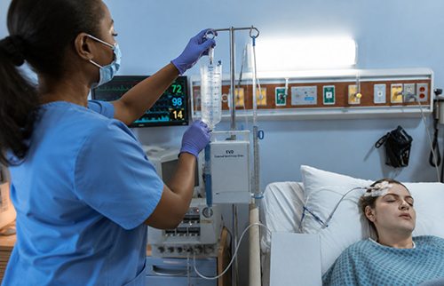
Common conditions that require an EVD drain include traumatic brain injury, bleeding around the brain (subarachnoid hemorrhage), and hydrocephalus. All of these involve dangerous levels of pressure in the skull.Common conditions that require an EVD drain include traumatic brain injury, bleeding around the brain (subarachnoid hemorrhage), and hydrocephalus. All of these involve dangerous levels of pressure in the skull.
Key EVD Drain Nursing Responsibilities
Managing an EVD drain involves several critical nursing tasks. Each one must be done correctly and consistently. Here is what nurses need to focus on:
- Level the EVD Drain at Every Shift - Always re-level the drain at the start of every shift and whenever the patient is repositioned. The drain should align with the foramen of Monro near the ear, as even small height changes can cause CSF to drain too fast or too slow, leading to serious complications or pressure buildup.
- Watch CSF Color and Amount - Check CSF hourly and document the amount drained. Normal CSF should be clear like water; if it appears cloudy, pink, or bloody, notify the doctor immediately, as this may indicate infection or bleeding. Both excessive and insufficient drainage can be dangerous.
- Keep the System Sterile -Because an EVD drain goes directly into the brain, strict sterile technique is essential. Never touch connection points without sterile gloves or flush the drain unless ordered by a doctor. Check the insertion site dailyfor redness, swelling, or discharge, and change the dressing using sterile technique according to hospital protocol.
- Read the ICP Waveform - When the EVD is also monitoring intracranial pressure (ICP), watch the waveform on the monitor. A healthy waveform has three peaks. When the middle peak rises higher than the first, that is a warning sign. It may mean the brain is losing its ability to manage pressure changes. Report this change to the care team right away.
- Clamp Before Transport - Always clamp the EVD drain before moving the patient. Forgetting this step is one of the most common and dangerous EVD errors. Use a checklist or ask a second nurse to verify before and after every transport. Once the patient is settled in the new position, unclamp the drain and re-level immediately.
Common EVD Drain Problems and What to Do
Even with great nursing care, EVD drains can have problems. Here is how to troubleshoot the most common ones:
- No drainage: Check that the drain is unclamped. Confirm the drain level isNo drainage: Check that the drain is unclamped. Confirm the drain level iscorrect. Look for kinks in the tubing. If drainage is still absent, call theneurosurgery team.
- Too much drainage: Verify the drain is set at the right level. If output isunusually high, notify the physician. Over drainage can cause a dangerous drop in brain pressure.
- Bloody CSF: Note the time and appearance. Document carefully. Do not flushthe drain. Contact the doctor immediately.
- Air in the tubing: Check all connections. Make sure they are tight. If a large air bubble is present, call the team before taking any action.
Building Your EVD Drain Skills With AANN
Managing an EVD drain well takes both knowledge and practice. The Core Curriculum for Neuroscience Nursing and the Evidence-Based Clinical Review: Intracranial Monitoring are some of the best resources available. It gives nurses a deep clinical foundation in neuroscience nursing, including intracranial pressure management.
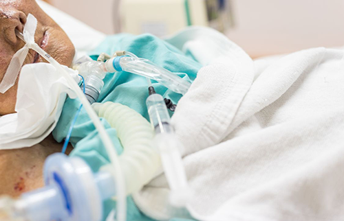
AANN also offers continuing education through its Annual Conference and online courses. These help nurses stay current with the latest evidence. Additionally, AANN membership connects you with a community of more than 5,200 neuroscience nurses.
Ready to Advance Your Neuroscience NursingReady to Advance Your Neuroscience Nursing Career?
EVD drain nursing is one of the most demanding skills in the specialty. It requires focus, clinical judgment, and a commitment to excellence. AANN supports nurses who take that commitment seriously. Become a member today and access the tools you need to deliver the best care possible.
Categories: Education | Professional Development | Neurotrauma
Standardizing Neurocheck Documentation for Improved Outcomes
In neuroscience nursing, the neurological check, or neurocheck, is one of the mostIn neuroscience nursing, the neurological check, or neurocheck, is one of the most important assessments a nurse performs. It helps detect early signs of brain or spinal cord changes. When done consistently and documented correctly, it can catch a problem before it becomes a crisis.
However, many nursing units lack a clear standard for when and how to perform these checks. The timing of neurocheck intervals varies from nurse to nurse and unit to unit.This inconsistency creates risk for patients.
Standardizing neurocheck intervals and documentation practices is one of the most effective steps a neuroscience team can take. In this article, we break down what a neurocheck includes, how often it should happen, and how to avoid the most common documentation mistakes.
What Does a Neurocheck Include?
A basic neurocheck measures how well the brain and nervous system are working. It is not complicated, but it must be done carefully and the same way every time. Key parts of a neurocheck include:
- Level of consciousness – Is the patient awake, alert, and responsive?
- Pupil check – Are the pupils equal in size? Do they react to light?
- Motor strength – Can the patient move their arms and legs equally on both sides?
- Speech and understanding – Can the patient speak clearly and follow instructions?
- Vital signs – Are blood pressure, heart rate, and breathing within normal limits?
Together, these checks paint a picture of the patient's current neurological status. The real value, however, comes from tracking changes over time. A small change in one area may not mean much alone. But a pattern of small changes can signal a serious problem.
Recommended Neurocheck Intervals: How Often Is Often Enough?
The right neurocheck interval depends on the patient's condition and how stable they are. There is no one-size-fits-all answer. However, there are general guidelines that most neuroscience units follow:
- Every 15 to 30 minutes – For patients just out of brain surgery, or thoseshowing signs of rapid change
- Every 1 to 2 hours – For patients with an active stroke, brain bleed, or traumatic brain injury
- Every 4 hours – For patients who are stable but still being monitored after a neurological event
- Every 8 to 12 hours – For patients in rehabilitation with stable, long-term neurological conditions
These neurocheck intervals should always be written in the patient's orders. The doctor or nurse practitioner sets the frequency. However, nurses must use their clinical judgment too. If something seems different, check the patient sooner. Do not wait forthe next scheduled interval.
For clinical practice guidance, the AANN Neuroscience Resources page is an excellent reference. It provides up-to-date tools for nurses working in any neuroscience setting.
Why Consistent Neurocheck Intervals Lead to Better Patient Outcomes
Consistency in neurocheck intervals is not just a policy issue. It directly affects patientsafety. When every nurse follows the same schedule and uses the same assessment tool, the documentation tells a clear story. The care team can then spot deterioration faster.
For example, consider a patient recovering from a stroke. Their left arm strength is 4/5 at 8 a.m. By noon, it drops to 3/5. By 2 p.m., it is 2/5. That trend is only visible when neurochecks are done consistently and documented precisely. Without that pattern, a worsening stroke may go unnoticed for hours.
Furthermore, consistent documentation protects nurses legally. It shows that proper monitoring was completed and any changes were reported in a timely way.
Common Neurocheck Documentation Mistakes to Avoid
Even skilled nurses can fall into habits that weaken the quality of their neurocheck documentation. Here are the most common mistakes and how to fix them:
- Copy-pasting the last assessment: This hides real changes. Every neurocheck must reflect a fresh, hands-on assessment, not a copied entry.
- Using vague terms: Words like 'oriented' or 'confused' mean different things to different people. Use a validated scale instead, such as the Glasgow Coma Scale(GCS).
- Skipping baseline comparisons: Always note whether findings are the same, better, or worse than the last check. Context matters.
- Late documentation: Record findings as soon as possible after the assessment. A long delay creates doubt about when a change actually occurred.
Building a Neurocheck Standard Across Your Unit
Standardizing neurocheck intervals requires teamwork. It is not enough to write apolicy. The whole unit must understand why consistent checks matter. Education, practice, and open communication are key.

Unit leaders can host brief case reviews to show staff how a missed or delayed neurocheck led to a poor patient outcome. These real-life examples are powerful teaching tools. They make the importance of the standard feel personal, not just procedural.
Additionally, neuroscience nurses can bring quality improvement ideas to the Neuroscience Nursing Annual Conference. Sharing what works, and what does not, strengthens the entire specialty.
How CNRN Certification Strengthens Neurocheck Skills
Nurses who earn the CNRN certification through the American Board of Neuroscience Nursing (ABNN) develop a strong foundation in neurological assessment. This includes neurocheck skills and documentation standards. Studying for the CNRN exam using the Core Curriculum for Neuroscience Nursing is one of the best ways to build this knowledge
The CNRN credential also shows employers and colleagues that a nurse has met a recognized standard of excellence. It is a mark of commitment to the specialty.
Join AANN and Elevate Your Neuroscience Nursing Practice
Neuroscience nursing is a demanding specialty. But you do not have to navigate it alone. AANN offers education, clinical resources, and a professional community to support your growth. Become a member today and connect with more than 5,200 nurses who are passionate about advancing the standard of care.
Categories: Education
Parkinson's Medication Timing and Mobility: What Neuroscience Nurses Need to Know
Consider the nurse who catches a patient mid-freeze at the bedside rail, an hour after a late dose. That moment is preventable. Parkinson's nursing care is built on one reality: the drug must arrive on time. Studies show that more than half of all Parkinson’s Disease (PD) doses during unplanned admissions arrive more than 30 minutes late. Even worse, nearly one-third are delayed for more than an hour. For patients whose motor function depends on steady dopamine levels, that gap causes real harm.
Why Timing Is Central to Excellent Parkinson's Nursing Care
PD destroys dopamine neurons in the substantia nigra. Carbidopa-levodopa replaces that signal. Yet the drug works only with consistent plasma levels. When doses fall outside the home schedule, patients shift into off states. Rigidity sets in. Gait freezes. Fall risk climbs. The NINDS describes PD as chronic and progressive. Indeed, each disruption compounds the next.
Three barriers disrupt inpatient dosing. First, rigid hospital schedules don't fit home dosing needs. Some patients need a dose every one to two hours. Second, units often don't stock the right drug forms. Third, dysphagia gets in the way when teams skip swallowing checks before the dose window opens.
Your admission assessment should address all three. Write down the home schedule in detail. Confirm drug availability with the pharmacy. Flag swallowing concerns for speech therapy before the first dose is due. Also, loop in the dietitian early. Protein competes with levodopa for absorption. Specifically, a high-protein meal near a dose can blunt the drug's effect and mimic an off state.
Monitoring Motor Fluctuations in Parkinson's Care
Motor fluctuations in Parkinson's nursing care follow a pattern you can track. On states bring mobility and less tremor. Conversely, off states bring rigidity, slow movement, and frozen gaits. Log these shifts against the dose schedule, as the comparison helps the team determine whether these reflect missed doses or disease progress. The answer shapes the care plan. It also helps the neurologist decide whether to adjust the dose or timing.
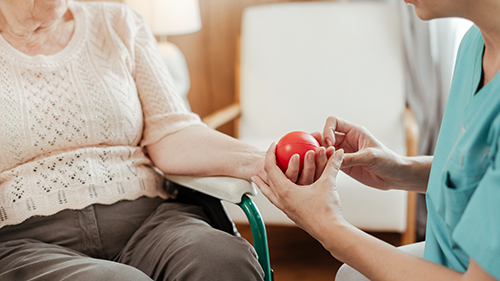
Standard fall-risk tools miss the mark in PD. They don't capture gait freezing, poor balance, or the sudden shift between on and off states. Instead, a focused check covers freezing episodes, trouble turning, and hazards in the room. Physical therapy early in the stay produces better results than a referral at discharge. So does early speech therapy. Aspiration events raise acuity, extend stays, and cut the activity that supports motor function.
A focused fall check includes the following:
Logging every freezing episode with time and trigger
Checking footwear, floor surfaces, and bed height on admission
Getting physical therapy involved within the first 24 hours
Teaching the patient and caregiver cueing strategies for frozen gait
Together, these steps cut the fall risk in ways that standard tools can miss entirely.
Strengthening Parkinson's Nursing Care Through Documentation
Good records anchor every handoff. Write down the drug form, exact dose times, and any notes from the neurologist. Research shows that nursing alerts in the chart improve on-time PD dosing. Furthermore, stocking the right drug forms in dispensing machines helps too. For example, units that used both saw fewer delayed doses and fewer off-state events. That evidence belongs in your unit's workflow.
Before discharge, confirm the outpatient schedule matches any changes made during the stay. Make sure the patient and caregiver can explain the dosing plan. Schedule a neurology follow-up appointment within a safe window. In addition, send home a written schedule in plain language. Teaching caregivers to spot off-period signs and manage frozen gait reduces readmission risk. Similarly, showing them how to respond to a freezing episode keeps the patient safer at home. These steps belong in every PD discharge plan.
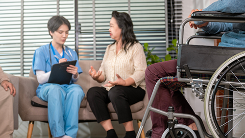
Ultimately, Parkinson's nursing care is what stands between a stable stay and a preventable decline. The field keeps moving, and keeping up matters at the bedside. The American Association of Neuroscience Nurses (AANN) supports nurses across the full PD care continuum. Parkinson's nursing care resources, mentorship, and professional growth tools are all part of membership. Additionally, the organization connects you with more than 5,200 colleagues who share that standard. Securing an AANN membership puts that community solidly behind your practice.
Frequently Asked Questions
What is the biggest medication risk for hospitalized Parkinson's patients? Delayed or missed carbidopa-levodopa doses are the most consequential risk. Even a 30-minute delay can push a patient into an off state, increasing rigidity, postural instability, and fall risk. Reconciling the home dosing schedule at admission and building it into the electronic medical record reduces timing errors during the stay.
How does dietary protein affect levodopa absorption? Large neutral amino acids from dietary protein compete with levodopa for intestinal absorption. Specifically, a high-protein meal close to a dose can blunt the medication's effect and produce symptoms that resemble an off state. Coordinating meal timing with the medication schedule, in collaboration with the dietitian, addresses this without changing the drug regimen.
Why do standard fall-risk tools underperform in Parkinson's patients? Generic fall-risk tools don't account for disease-specific mechanisms: freezing of gait, poor balance, and the abrupt shift between on and off states. A targeted assessment that documents freezing episodes, trouble turning, and room hazards provides an accurate picture of risk.
What should a nurse confirm before discharging a Parkinson's patient? First, confirm if the outpatient schedule matches any inpatient changes. Then, check if the patient and caregiver can explain the dosing plan. Finally, check if a neurology follow-up is scheduled. Teaching caregivers to spot off-period signs and manage freezing at home is a direct readmission prevention measure.
Categories: Parkinsons | Movement Disorders | Education
Hemorrhagic Stroke Nursing Care: Managing Acute Intracerebral Hemorrhage (ICH)
A patient arrives with a sudden bad headache and a dropping GCS score. After imaging confirms a large bleed in the brain, what happens in the next hour will determine the patient’s outcome. An attack of hemorrhagic stroke carries a 30-day death rate near 40%, with the bulk of the risk occurring in the first hours.
Most intracerebral hemorrhage (ICH) deaths due to hemorrhagic stroke happen during the first hours. The nurse running serial neuro checks, titrating the drip, and flagging the one-point GCS drop is the reason some patients don't become that statistic.
Blood Pressure Control in Hemorrhagic Stroke Care
Hematoma growth affects roughly one-third of ICH patients in the first 24 hours. It's one of the strongest signs of coming decline and death. The 2022 AHA/ASA guideline for ICH recommends starting blood pressure treatment within two hours of onset. For patients with systolic blood pressure between 150 and 220 mmHg, the target is 140 mmHg. Reaching that target within one hour is the standard.
Blood pressure readings turn inaccurate when the applied technique is inconsistent. Cuff size, limb position, patient agitation, and outside stimulation each can skew the number. A reading that looks controlled may simply reflect a bad measurement, so document the method alongside the value every time. When readings climb despite protocol compliance, escalate the situation immediately. Chances are that the next scheduled chart check might be too late.
The same guideline recommends care in an ICU or stroke unit with neuroscience-trained nurses. Continuous nursing checks of intracranial pressure (ICP), cerebral perfusion pressure (CPP), and blood flow help improve outcomes in this group.
Neuro Checks and ICP Tracking in Hemorrhagic Stroke
Serial neuro checks are the backbone of ICH care. Glasgow Coma Scale (GCS) scores and pupil checks track the impact of bleed volume, brain swelling, and fluid buildup in the patient. All of these can shift fast in the first 48 to 72 hours. A one-point GCS drop, new pupil asymmetry, or Cushing's triad warrants immediate escalation. Waiting for the next scheduled check when these signs are already visible isn’t what you would call a safe environment.
Elevated ICP causes harm through several paths. Head-of-bed elevation at 30 degrees reduces venous outflow resistance. Hyperosmolar therapy requires osmolality checks and fluid balance. Temperature control carries risks around shivering and sedation depth. Still, sedation must not mask the neuro exam. That exam is your earliest warning. You can't afford to lose it.
ICH also raises the risk of cortical seizures. Non-convulsive activity can happen without visible motor signs. When nurses order continuous EEG monitoring, those who know the clinical reason maintain lead contact and report findings clearly. Indeed, an NCBI overview of hemorrhagic stroke confirms that nurses drive early decline detection in ICH. That role needs both clinical skill and unit-level practice.
Managing Problems and Planning Discharge in Hemorrhagic Stroke
ICH brings several serious problems: bleeding extension into the ventricles, fluid buildup requiring an external ventricular drain (EVD), and clotting issues needing reversal. EVD care, whether it’s zeroing, leveling, and troubleshooting, requires training that goes beyond general ICU orientation. A nurse managing a drain needs to know what a waveform change means and when to call. Structured neuroscience education builds those skills in a way that on-the-job learning alone does not.
Goals-of-care talks are also a core part of hemorrhagic stroke nursing. Prognosis often reaches families within hours of the bleed. Nurses are frequently the most steady presence when families process that news over subsequent shifts. Accurate updates on what the team sees, what they're doing, and what they're watching for next is direct care. It's not a side task.
Discharge planning in ICH starts early. Rehab potential, caregiver support, and the patient's likely path all shape placement choices. Nursing checks inform those choices. Furthermore, good handoffs reduce the risk of gaps between acute care and rehab. Ultimately, the quality of care after the unit depends on the groundwork laid before discharge.
Hemorrhagic stroke nursing care is built on speed, skill, and steady presence at the bedside. AANN offers the Seizure and Epilepsy Certificate Program, clinical tools, and community support that this work demands. In addition, getting an AANN membership connects you with more than 5,200 nurses who share that focus.
Frequently Asked Questions
What is the blood pressure target for acute intracerebral hemorrhage? The 2022 AHA/ASA guideline recommends starting blood pressure treatment within 2 hours of ICH onset and reaching the target within 1 hour. Meanwhile, for patients with systolic blood pressure between 150 and 220 mmHg, the target is 140 mmHg. Smooth, steady control that avoids spikes remains the standard.
What neuro changes in ICH require immediate escalation? A one-point GCS drop, new pupil asymmetry, or Cushing's triad each warrants immediate escalation. These signs can reflect hematoma growth, fluid buildup, or rising ICP. All require fast action to limit further harm. Waiting for the next scheduled check when these signs appear isn’t advisable.
Why does ICH care require neuroscience-trained nurses specifically? The 2022 AHA/ASA guideline recommends care in an ICU or stroke unit with neuroscience-trained nurses. Continuous checks of ICP, CPP, and blood flow, combined with the ability to catch subtle neuro changes, require specialized skill. General inpatient training doesn't reliably build that foundation.
What problems should nurses watch for in ICH patients? Bleed extension into the ventricles, fluid buildup requiring an EVD, clotting issues needing reversal, and cortical seizures, including non-convulsive activity, are the main concerns. Each needs specific skills: EVD care, osmolality checks, clotting status tracking, and EEG monitoring, where indicated.
From Brain Anatomy to Seizure Management: Mastering Neuro Assessment and Epilepsy Care
A patient who was chatting an hour ago is now hard to rouse. One pupil reacts a little slower than the other. Caught early, those two findings can be the first sign of a brain in trouble. Neurological assessment (also known as neuro assessment) in nursing is the disciplined skill of noticing them and acting before a small change becomes a crisis.
The work is methodical: checking consciousness, pupils, and movement, then tracking how the findings shift. When caring for someone with seizures or epilepsy, that vigilance is anything but routine. It often decides whether the team catches a deficit in time.
The Building Blocks of Neuro Assessment
A reliable exam rests on a few core components, done the same way every time and recorded clearly:
Level of consciousness, often scored with the Glasgow Coma Scale (GCS)
Pupil size, equality, and reactivity to light
Motor strength and symmetry in all four limbs
Speech, orientation, and cranial nerve function
Vital sign trends that can signal rising intracranial pressure
Each component tells part of the story, and a change in any one can be the earliest clue. The real value, however, lies in repetition. A single check is a snapshot, but a series reveals the trajectory that drives every decision after it. Yet an exam helps only if the team can act on it. Clear, consistent notes allow the next clinician to compare findings and spot trends across the shift.
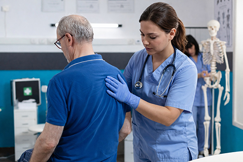
A structured handoff matters just as much. The strongest ones include the specific finding, when it appeared, and the change since the last check. For example, a two-point drop in the GCS prompts action in a way that "looks worse" never will. The American Association of Neuroscience Nurses’ (AANN) clinical practice guidelines give clinicians a shared, evidence-based reference for performing and standardizing these exams.
Applying the Exam in Seizures and Epilepsy
Epilepsy is common. About 2.9 million U.S. adults live with active epilepsy, roughly one percent of the adult population. So good care depends on what the bedside team sees before, during, and after a seizure. Some patients sense a warning aura beforehand, which buys time to prepare.
During an event, the team records the start time, the duration, and the movement pattern. Recognizing the seizure type, from focal to generalized, shapes both the workup and the response. Still, the exam afterward is just as important. A long delay in waking, a new one-sided weakness, or a seizure that will not stop signals an emergency.
Status epilepticus is one such emergency. It means a seizure that lasts five minutes or longer, or repeated seizures without a return to baseline between them. A sharp eye is what catches it early. The warning signs include:
A convulsive seizure that lasts for more than five minutes
Repeated episodes with no recovery between them
Subtle signs of ongoing activity, such as eye deviation or rhythmic twitching, in someone who is not waking up
Together, fast recognition, airway protection, and timely treatment protect the brain from lasting harm. Recording the exact times is critical because every later decision depends on them.
Strengthening Neuro Assessment Through Nursing Education
Skill at the bedside grows with structured learning. For clinicians who care for this population, the AANN/ABNN Seizure and Epilepsy Certificate Program builds focused, practical knowledge. It covers seizure types, monitoring, and care in the epilepsy monitoring unit.

Quick-reference tools can also help in the moment. AANN's neuroscience resources also include checklists and guides that reinforce a consistent routine, which helps those newer to neuro. Formal courses and quick guides each shorten the path from competent to expert.
Sharpen the Skills Your Patients Depend On
A strong neurological exam is the quiet foundation of safe neuroscience care. That skill sharpens with every repetition and every new piece of evidence. So if you want to build that expertise alongside a community of fellow specialists, consider availing of an AANN membership. It connects you to the education, guidelines, and colleagues that support your growth.
Frequently Asked Questions
What is the most important part of a neurological exam? No single piece stands alone, but the level of consciousness is often the most sensitive early sign of change. A drop in the Glasgow Coma Scale score frequently shows up before anything else. Still, the most reliable read comes from assessing all the components together and comparing them to the patient's last exam.
How often should neuro checks be done? It depends on the patient's condition, diagnosis, and unit protocol. A stable patient may need a check every few hours. An unstable or post-procedure patient may need one every fifteen minutes to an hour. Consistency is the key, so any change is caught quickly and recorded clearly.
What should a nurse do right after a patient has a seizure? First, secure airway, breathing, and safety. Then complete a focused exam, noting how long the seizure lasted and how quickly the patient returns to baseline. Report any prolonged recovery, new deficit, or repeated event promptly, since these can signal a need for urgent care.
How can a nurse build stronger neuro assessment skills? Practice consistently, use a standardized framework, and learn from current guidelines and structured education. Certificate programs and specialty resources for neuroscience and epilepsy can speed that growth, especially with feedback from experienced colleagues.
Investing in Excellence: Strategies and Events for Nursing Professional Development in Neuroscience
Consider the clinician who can manage an external ventricular drain, recognize status epilepticus, and read a subtle pupillary change. None of these learnings can be acquired from reading a textbook alone. Instead, these skills take years of steady nursing professional development, the type of training that turns a generalist into a confident specialist.
Meanwhile, advances in fields such as stroke protocols, seizure care, and neurocritical care continue to flourish. Each September, Nursing Professional Development Week marks that commitment to continuous improvement. For the neuroscience team, it’s also a good prompt for a simple question: What’s your development plan this year?
Why Nursing Professional Development Matters in Neuroscience
The case for learning more is practical, not abstract. For example, research links continuing professional development to stronger knowledge, sharper skills, and better patient outcomes. In neuroscience, a patient's examination can change in minutes, so current knowledge has direct value at the bedside. A nurse who studied the latest stroke guidelines is quicker to catch an early decline and act sooner. Development also protects the workforce. Nurses who feel they are advancing their skills report more job satisfaction and tend to stay longer at their jobs.
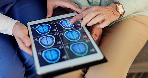
Build a Nursing Professional Development Plan
Learning works best when it follows a specific direction. First, look at the numbers. Employment of registered nurses is projected to grow 5 percent from 2024 to 2034, faster than average. In a field this large, your specialty is what sets you apart.
Before you sign up for the next course, map the year against where you want to go. A focused plan might include:
A target credential or certificate program tied to your specialty
One major educational event for depth and networking
A defined clinical skill to build, with a way to measure progress
A mentor relationship to guide bigger decisions
Ultimately, writing the plan down turns a vague intention into a schedule you can follow. A clear goal is also easier to fund when you ask a manager for support.
Events and Education That Move You Forward
The American Association of Neuroscience Nurses (AANN) offers several ways to build expertise throughout the year. The Annual Conference, for example, brings the specialty together for deep learning, leadership sessions, and peer connection. In addition, shorter courses and webinars fit continuing education into a busy schedule, with no travel. Each one carries CE contact hours that count toward your license and recertification. For nurses who learn best through people, AANN's mentorship program pairs members with experienced colleagues. A mentor can help you weigh a first charge-nurse role or a move into advanced practice.
For many neuroscience nurses, the next milestone is a formal credential. Certification confirms a broad body of specialty knowledge through an exam. The CNRN® and SCRN® credentials from the American Board of Neuroscience Nursing are two examples. In contrast, a certificate program offers focused training in one area, such as epilepsy or spine care. Both belong in a development plan. Each one gives a year of study a clear, recognized goal, and naming that target early shapes the rest.
Nursing Professional Development Requires You to Make Learning a Habit and Not an Event
The nurses who advance the most treat development as continuous, not occasional. They debrief tough cases, read current guidelines, and bring questions to colleagues. A conference gives you a burst of learning, but a monthly habit is what adds up. So set a recurring block each month to review one study or finish one short module. Over a year, those small steps become real progress. Also, a few minutes spent noting what a hard case taught you turns experience into lasting knowledge.

That kind of development turns daily neuroscience experience into a recognized, lasting skill. The field will keep changing, and a plan is how you stay ahead of it. So if you want a community behind that plan, consider investing in an AANN membership. It connects you to the education, events, and network that keep you moving forward.
Frequently Asked Questions:
How is professional development different from continuing education credits? Continuing education credits are one part of professional development, often required to keep a license or credential. However, professional development is broader. For example, it includes mentorship, leadership training, conferences, and self-directed learning that build a career over time, not just the hours logged for compliance.
How much professional development should a neuroscience nurse aim for each year? There is no single number that fits every role. Instead, set goals tied to your career direction rather than a minimum hour count, then choose activities that move you toward them. Many nurses combine one larger event with smaller, regular learning throughout the year.
Does professional development really affect patient care? Research links continuing professional development to stronger knowledge and skills, and in many studies, to better patient outcomes. Still, the effect depends on how well the learning is designed and applied. In neuroscience, staying current with new assessment and treatment standards directly supports safer care.
What is the best first step for a nurse new to neuroscience? First, pair foundational learning with a mentor. Core neuroscience resources build a base, while an experienced colleague helps you apply that knowledge and avoid common mistakes. From there, a specialty credential or certificate program becomes a natural next goal.
Categories: Professional Development | Leadership
Bridging the Education Gap: Empowering the Neuroscience Advanced Practice Provider With a New Certificate Program
As neuroscience care grows more complex, the demand for specialized knowledge among advanced practice providers has never been higher. Neuroscience nurse practitioners and other advanced practice providers (APPs) are stepping into roles that require deep clinical expertise, yet formal neuroscience training during graduate education often falls short of what practice demands.
A 2025 study published in Neurology Education found that 97.6% of nurse practitioner programs include neuroscience didactics. It also found that only 18% cover lesion localization and 24.3% cover neuroradiology. Meanwhile, only 82% of programs offer a neurology clinical rotation. Of those, approximately 77.5% report that 10% or fewer students complete the rotation each year. The single most commonly cited barrier is a lack of neurology preceptors.
These gaps leave many APPs entering neuroscience practice underprepared, making targeted post-graduate education not just valuable, but essential.
The Expanding Role of Neuroscience Nurse Practitioners
Advanced practice providers play a critical and growing role in neuroscience patient care and system-level leadership. A recent AANN survey found that the practice landscape of neuroscience APPs across the United States remains largely understudied. Findings from that survey underscore the urgent need for standardized measures of neuroscience nurse practitioners.
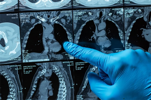
The scope of neuroscience APP practice spans the full continuum of care, including inpatient and outpatient settings and geriatric neurology. This breadth demands a structured, evidence-based educational foundation. Without it, newer APPs face a steep learning curve that can affect both provider confidence and patient outcomes.
Certificate Program for the Seizure and Epilepsy Healthcare Professional
The value of structured certificate education has already been demonstrated. A 2025 prospective cohort study published in the Journal of Neuroscience Nursing examined the effects of the AANN Seizure and Epilepsy Certificate Program on 39 neuroscience nurses in an epilepsy monitoring unit (EMU). Results were compelling:
Mean epilepsy knowledge scores rose from 12.3 to 16.1 (P < .001)
Self-reported confidence scores increased from 19.0 to 22.1 (P < .001)
Time from seizure identification to treatment decreased from a mean of 6.5 minutes to 4 minutes
The study also found that improvements in knowledge correlated with the highest terminal degree attained, reinforcing the principle that advanced-level learners benefit most from advanced-level curricula. These outcomes affirm that certificate programs can move the needle on both provider competency and real-world patient safety.
What the New Neuroscience APP Certificate Program Offers
Responding directly to the documented education gap, AANN and the American Board of Neuroscience Nursing (ABNN) are currently developing the Neuroscience Advanced Practice Provider (APP) Certificate Program. This assessment-based, online self-study program is specifically designed for newer APPs or APPs new to neuroscience who need a broad curriculum covering topics across the care continuum.
The program's general learning outcomes reflect a high clinical standard:
Demonstrate a comprehensive understanding of pathophysiology, clinical presentation, and management strategies across a broad range of neurological disorders
Exhibit proficiency with advanced diagnostic tools for diagnosing neurologic conditions
Coordinate comprehensive care integrating multidisciplinary approaches in both inpatient and outpatient settings
Exhibit leadership in ethical decision-making, informed consent, and end-of-life care planning
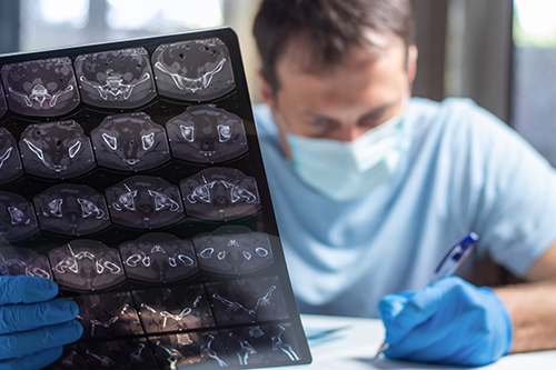
The curriculum spans 13 content modules, covering everything from foundations of neuroscience and cerebrovascular conditions to neurocritical care, epilepsy, spine disorders, neuromuscular disorders, geriatric neurology, and advanced practice ethical and legal considerations. Learners who successfully complete the program and pass the post-assessment will earn continuing education (CE) contact hours and an ABNN certificate of added qualification.
Spine Care and a Growing Certificate Portfolio
The AANN and ABNN certificate portfolio is expanding beyond the Neuro-APP Certificate Program. The Certificate Program for the Spine Healthcare Professional is an eight-module, assessment-based online course designed to provide foundational knowledge and evidence-based care for patients with spinal disease or injury requiring medical or surgical treatment. Participants who complete all eight modules and pass the post-assessments will also earn CE contact hours and an ABNN certificate of added qualification.
Together with the existing Seizure and Epilepsy Certificate Program, these offerings represent a deliberate strategy: building a ladder of specialized, credentials-backed education that meets nurses and APPs wherever they are in their career trajectory. Each program uses instructional design principles to deliver structured online learning followed by post-assessment, ensuring that earning a certificate reflects genuine knowledge attainment rather than participation alone.
Take the Next Step in Your Neuroscience Practice
If you are an advanced practice provider working in neuroscience, now is the time to formalize the expertise you bring to your patients every day. AANN's and ABNN’s growing suite of certificate programs offers a flexible, rigorous, and clinically relevant path to do exactly that. Whether you are new to neuro practice or looking to deepen your competency in a specific domain like epilepsy or spine care, there is a program designed for you.
Visit the Certificate Programs page of the website to learn more.
FAQ
Q: Who is eligible to enroll in the Neuroscience APP Certificate Program?
A: The program targets newer advanced practice providers, or APPs who are new to neuroscience, who are seeking a comprehensive, broad neuroscience curriculum across the care continuum. Nurse practitioners, clinical nurse specialists, and physician assistants working in neuro settings are all appropriate candidates.
Q: Is the ABNN certificate of added qualification the same as board certification?
A: No. A certificate of added qualification is earned through a structured, assessment-based educational program and indicates attainment of specific knowledge. It is distinct from board certification, which involves a separate eligibility and examination process through ABNN.
Q: Do these certificate programs meet any accreditation requirements?
A: Yes. The Seizure and Epilepsy Certificate Program is designed to help facilities meet National Association of Epilepsy Centers (NAEC) accreditation requirements for nursing expertise in epilepsy, particularly for nurses working in a comprehensive epilepsy center or epilepsy monitoring unit.
Q: How long does it take to complete a certificate program?
A: Each program is self-paced and completed online. The number of modules varies by program. For example, the Seizure and Epilepsy program includes six required modules plus two optional supplementary modules, while the Spine program consists of eight sequential modules with a post-test.
Q: Can completing a certificate program replace a neurology clinical rotation?
A: No. Certificate programs are designed to supplement and strengthen post-graduate clinical knowledge, not replace experiential training. They serve as a rigorous educational foundation that supports evidence-based practice in specialty neuroscience settings.
Categories: Professional Development | Advanced Practice
Strategies for Success as a New Stroke Coordinator
Stepping into the stroke coordinator role is both an exciting and demanding career transition. The position carries a broad scope of clinical, administrative, and educational responsibilities that can feel overwhelming in the early months. With the right strategies and support, however, new stroke coordinators can build a strong foundation and lead programs that deliver measurable improvements in patient outcomes.
Understanding the Full Scope of the Role
The stroke coordinator role is often described as the "glue that holds the program together." It encompasses clinical consultation, quality data management, staff education, community outreach, and regulatory compliance. These responsibilities span multiple departments and require building trust with physicians, nurses, and allied health professionals simultaneously.

One of the most common challenges new stroke coordinators face is not fully understanding the depth of this role from the start. Research shows that unclear role expectations are a leading contributor to early burnout and turnover among stroke coordinators. Taking time in your first weeks to map out your organization's specific expectations is a critical first step.
Prioritizing Data Literacy and Regulatory Knowledge
Data management is at the core of stroke program success. Stroke coordinators are responsible for abstracting and entering data into registries such as Get With The Guidelines (GWTG-Stroke), analyzing quality metrics, and presenting findings to medical leadership. Proficiency with these tools is non-negotiable.
New coordinators should prioritize orientation to GWTG-Stroke and make time to work alongside data abstraction teams early in their tenure. Regulatory literacy is equally important. Understanding The Joint Commission (TJC) stroke certification standards and how they translate into daily practice helps new coordinators anticipate audit requirements and lead program compliance with confidence.
Key early data and compliance priorities include:
Learning the GWTG-Stroke platform and completing data entry with accuracy
Reviewing concurrent chart documentation to ensure clinical pathway adherence
Familiarizing yourself with TJC stroke-specific standards and quality measures
Building Interdisciplinary Relationships
A stroke coordinator cannot succeed in isolation. The role demands consistent collaboration with emergency medicine, neurology, neurosurgery, vascular surgery, rehabilitation, and nursing leadership. Relationships built early tend to make later conversations about process improvement far more productive.

Facilitating interdisciplinary stroke team meetings and rounding on stroke patients with a multidisciplinary lens positions the coordinator as both a clinical resource and a program advocate. Communication skills, both verbal and written, are foundational. The ability to translate clinical data into actionable insights is what elevates a good coordinator to a great one.
Investing in Education and Mentorship
Staff education is a formal part of the stroke coordinator's responsibilities, but ongoing self-education is equally essential. Formal preparation programs, including stroke boot camps and structured onboarding pathways, have been shown to significantly reduce role confusion and improve coordinator retention.
Mentorship is one of the most powerful tools available. In a survey of stroke coordinators, 13 out of 14 respondents said mentorship would have helped their onboarding significantly, and all 14 said knowing their roles and responsibilities clearly from day one would have eased the transition. Seeking out a mentor, whether through your health system, regional networks, or professional communities, accelerates both clinical growth and program performance.
Consider these professional development actions as a new coordinator:
Pursue the Stroke Certified Registered Nurse (SCRN) credential to deepen clinical knowledge
Attend stroke sessions at nursing conferences to stay current on evolving evidence-based guidelines
Identify a mentor with stroke program experience for structured guidance
Engage with EMS partners and community education programs from the outset
Grow Your Practice With AANN
If you are navigating the early stages of your stroke coordinator career, you do not have to do it alone. AANN offers targeted education and resources designed specifically for stroke nurses and coordinators. The Annual Conference and Advances in Stroke Care Conference feature sessions focused on the evolving stroke coordinator role, program management tactics, and data integration strategies.
AANN also provides SCRN preparation materials and access to the NeuroNetwork, where you can connect with other healthcare professionals about stroke-related topics. Investing in yourprofessional development through AANN is a direct investment in your patients, your team, and your program's long-term success.
Join AANN for access to stroke nursing resources and to connect with other stroke care professionals.
Frequently Asked Questions
What is the difference between a stroke coordinator and a stroke navigator?
A stroke coordinator typically manages administrative responsibilities such as certification requirements, data reporting, and regulatory compliance. A stroke navigator focuses more on patient interaction, follow-up care, and interdisciplinary coordination at the bedside. Many programs use both roles to divide responsibilities and improve overall program outcomes.
Do I need a specific certification to become a stroke coordinator?
There is no single required credential, but many stroke coordinators pursue the Stroke Certified Registered Nurse (SCRN) certificate program to validate clinical expertise.
How long does onboarding typically take for a new stroke coordinator?
Onboarding timelines vary by institution, but research suggests that formalized onboarding with mentorship, role clarity, and regulatory orientation leads to better outcomes. Many coordinators report that a structured 90-day orientation that includes data management training, chart review practice, and interdisciplinary introductions sets a strong foundation.
What are the biggest challenges new stroke coordinators face?
Common challenges include not fully understanding the breadth of the role, limited peer support, insufficient time to complete all responsibilities, and inadequate resources. Seeking mentorship and proactively establishing relationships with your medical director and hospital leadership can help address these issues early.
Categories: Education | Stroke | Professional Development
A Practical Guide to Grading Subarachnoid Hemorrhage
Subarachnoid hemorrhage (SAH) is a neurological emergency with mortality rates that can exceed 40%. Prompt and accurate grading is critical for guiding clinical decisions. The Fisher Scale remains one of the most widely used radiographic grading tools in SAH management. It helps clinicians predict the risk of cerebral vasospasm, one of the most feared complications following aneurysmal SAH.
Understanding how grading scales work is essential for neuroscience and stroke nurses. Knowing how to apply them in acute care settings supports faster decision-making and clearer interdisciplinary communication.
Understanding Subarachnoid Hemorrhage and Why Grading Matters
SAH occurs when blood enters the subarachnoid space, most often due to a ruptured cerebral aneurysm. The volume and distribution of blood visible on imaging are strong predictors of secondary injury. Vasospasm, delayed cerebral ischemia, and hydrocephalus are all associated with the extent of hemorrhage. Grading systems give clinicians a standardized way to assess severity and communicate risk across care teams.

A consistent grading language supports earlier intervention and more coordinated care. This is especially critical in high-volume stroke and neuroscience centers where rapid triage is the norm. Standardized grading also enables more meaningful outcome comparisons across institutions and research studies.
The Fisher Scale: Radiographic Classification of SAH
Introduced in 1980, the original Fisher Scale classifies SAH based on blood appearance on non-contrast CT imaging. It assigns patients to one of four grades:
Grade 1: No blood detected on CT
2: Diffuse or vertical layers of subarachnoid blood less than 1 mm thick
3: Localized clot or vertical layer of blood 1 mm or greater in thickness
4: Intracerebral or intraventricular clot, with or without diffuse subarachnoid blood
Grade 3 carries the highest predicted risk of vasospasm in the original scale. Experts designed the Fisher Scale to predict symptomatic vasospasm, not overall clinical severity. It does not account for the patient's neurological status at presentation. This distinction is important when integrating the scale into clinical workflows alongside other assessment tools.
The Modified Fisher Scale and Complementary Grading Tools
The Modified Fisher Scale, developed by Claassen and colleagues in 2001, addressed limitations of the original. It incorporated the presence of intraventricular hemorrhage and revised the classification of blood distribution. Research has shown that the Modified Fisher Scale improves predictive accuracy for delayed cerebral ischemia.
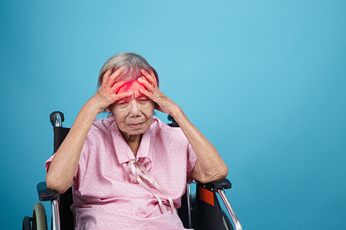
Other scales serve complementary clinical purposes:
Hunt and Hess Scale: Grades neurological status at presentation from Grade I (mild headache, no neurological deficit) to Grade V (deep coma, decerebrate posturing). It is widely used to assess surgical risk and predict overall outcome.
World Federation of Neurological Surgeons (WFNS) Scale: Combines the Glasgow Coma Scale score with the presence or absence of a motor deficit. It is considered more objective and reproducible than the Hunt and Hess Scale.
Clinical Implications for Neuroscience Nursing Practice
Grading scales are not merely documentation tools. They directly inform nursing priorities and patient monitoring plans. A patient with a Modified Fisher Grade 3 or 4 requires vigilant neurological monitoring for signs of vasospasm. Vasospasm typically peaks between days 4 and 14 after the initial bleed. Early detection of neurological change can be the difference between timely intervention and permanent deficit.
Neuroscience and stroke nurses are often the first to detect subtle changes in a patient's neurological status. A working knowledge of SAH grading helps nurses contextualize those observations. It supports more effective handoff communication, earlier escalation, and stronger collaboration with the neurosurgical team. Grading literacy is a professional competency that extends well beyond the physician's role.
Nurses who can accurately interpret grading findings position themselves better to advocate for their patients. This includes communicating clinical changes in a language that resonates with the entire care team. It also means recognizing when a patient's trajectory does not align with their grade. This discrepancy may signal a complication requiring immediate attention.
Advance Your Expertise in Neuroscience Nursing
Staying current on clinical tools like SAH grading scales is part of delivering high-quality, evidence-based care. The American Association of Neuroscience Nurses (AANN) provides continuing education and stroke-focused resources to help support your practice.
Learn more about the Stroke Certified Registered Nurse (SCRN) credential. The SCRN credential formally recognizes the attainment and demonstration of a unique body of knowledge necessary for the practice of stroke nursing.
Frequently Asked Questions
What is the difference between the Fisher Scale and the Hunt and Hess Scale? The Fisher Scale is a radiographic tool based on CT imaging findings, specifically designed to predict the risk of vasospasm. The Hunt and Hess Scale assesses the patient’s neurological status at presentation and predicts surgical risk and overall outcome. The two scales measure different dimensions of SAH severity and clinicians typically use them together to obtain a more complete picture.
Is the Modified Fisher Scale replacing the original Fisher Scale in clinical practice? The Modified Fisher Scale is increasingly preferred in research and many clinical settings because of its improved predictive accuracy for delayed cerebral ischemia. However, the original Fisher Scale remains in common use. Institutional protocols vary, so familiarity with both versions is valuable in practice.
Can a patient have a high Fisher Scale grade but a mild clinical presentation? Yes. The Fisher Scale reflects radiographic findings only. A patient may have a large clot burden on CT but present with minimal neurological deficits. Conversely, a patient with a low Fisher grade may still have significant clinical impairment. This is precisely why combining the Fisher Scale with a clinical grading tool like the WFNS Scale is important.
What is delayed cerebral ischemia and how does it differ from vasospasm? Vasospasm refers to the narrowing of cerebral arteries following SAH, which can reduce blood flow to the brain. Delayed cerebral ischemia (DCI) is a broader term that describes the clinical deterioration resulting from that reduced perfusion. Not all patients with radiographic vasospasm develop DCI, and some develop DCI without documented vasospasm. The Modified Fisher Scale was developed specifically to improve prediction of DCI risk.
Why Silent Strokes Are a Significant Risk for Future Infarcts
troke is one of the most studied neurological events in clinical practice. Yet a substantial threat often goes undetected until it appears on an imaging report. Silent stroke symptoms are so subtle that most patients never recognize them. Silent cerebral infarcts (SCIs) occur without obvious neurological deficits. However, evidence consistently shows they are not benign findings. They carry measurable, well-documented risk for future symptomatic infarction.
Understanding the Pathophysiology of Silent Stroke Symptoms
Silent strokes share the same underlying mechanisms as clinically apparent strokes. Small vessel occlusion, arteriolosclerosis, and thromboembolic events are all implicated. These infarcts most commonly occur in subcortical structures, including the basal ganglia, thalamus, and deep white matter. These regions do not govern highly visible motor or language functions. As a result, acute deficits go unnoticed.

Studies estimate that SCIs are present in approximately 10% to 38% of older adults. Prevalence varies based on imaging modality and population characteristics. Magnetic resonance imaging, particularly diffusion‑weighted sequences, detects these lesions far more sensitively than CT. Clinicians often identify many of the lesions incidentally during workup for unrelated neurological concerns.
The underlying small vessel pathology is progressive. Each new infarct adds to cumulative neural network disruption. This trajectory does not plateau. It accelerates.
Recognizing the Subtle Clinical Presentation
The term "silent" is accurate in one sense. There is no acute stroke presentation. Yet patients often do experience changes over time. These changes are gradual and are frequently attributed to aging or other conditions.
Clinicians assessing patients with known vascular risk factors should take note of the following:
Mild short-term memory lapses or reduced cognitive processing speed not explained by other causes
Subtle gait changes, postural instability, or a decline in fine motor coordination
New onset depression, emotional lability, or unexplained fatigue
Difficulty with executive function tasks that were previously manageable
None of these findings confirms an SCI. However, in a patient with a history of hypertension, diabetes, or atrial fibrillation, they demand a structured neurological evaluation. A comprehensive assessment can reveal functional deficits that standard intake screening will miss entirely.
The Evidence Linking Silent Stroke Symptoms to Future Risk
Silent cerebral infarcts are independent predictors of future symptomatic stroke. Data from multiple large longitudinal cohort studies demonstrate a two- to three-fold increase in stroke risk among individuals with SCIs compared to those without them. This elevated risk persists even after controlling for traditional vascular risk factors.
The Cardiovascular Health Study produced particularly compelling findings. SCIs detected on brain MRI were significantly associated with incident symptomatic stroke over time. The relationship held across sex, age groups, and varying degrees of white matter disease. Additional research has linked SCIs to accelerated cognitive decline and increased risk of vascular dementia.

For clinicians in acute, critical care, and rehabilitation settings, these findings carry direct clinical weight. A patient admitted for a first symptomatic stroke may already have a burden of silent infarction. That history shapes prognosis. It should inform care planning from admission onward.
Risk Factor Identification and the Nursing Role
Early identification begins with understanding which patients carry the highest burden of vascular risk. The following conditions are most strongly associated with silent infarct development and recurrence:
Uncontrolled hypertension, the single most modifiable contributing factor
Poorly managed type 2 diabetes mellitus
Atrial fibrillation and other cardioembolic sources
Hyperlipidemia and metabolic syndrome
Obstructive sleep apnea
Active tobacco use
Nurses are uniquely positioned to identify these patients before a symptomatic event occurs. Thorough neurological assessments, detailed vascular history documentation, and structured patient education are all within the nursing scope of practice.
Commit to the Knowledge Your Patients Need
If you care for patients at risk for stroke, the silent cerebral infarct is not a peripheral topic. It is central to how we understand and intervene in the full stroke continuum.
AANN provides continuing education and resources specifically designed to keep neuroscience nurses and advanced practice providers at the leading edge of clinical practice. Our stroke-focused professional development content is grounded in current evidence and built for the complexity of your clinical environment.
Learn more about AANN membership here.
Frequently Asked Questions
Can a patient have multiple silent strokes without ever having a symptomatic one? Yes. Some individuals accumulate a significant burden of SCIs visible on neuroimaging without ever presenting with a clinically recognized stroke. This pattern is particularly common in patients with longstanding uncontrolled hypertension and advanced small vessel disease. The absence of a symptomatic event does not indicate an absence of ongoing cerebrovascular injury.
Is there a standard screening protocol for silent strokes in high-risk populations? There is currently no universal screening recommendation for asymptomatic SCIs in the general population. However, several professional and cardiovascular societies recommend brain MRI consideration in patients with atrial fibrillation, recurrent unexplained cognitive symptoms, or high cardiovascular risk burden. Clinical judgment and individual patient context guide imaging decisions.
How do silent strokes affect a patient's rehabilitation potential after a symptomatic stroke? A pre-existing burden of SCIs is associated with reduced neuroplasticity and a more limited response to rehabilitation interventions. Patients with multiple SCIs prior to a symptomatic infarct may exhibit slower functional recovery and are at greater risk for post-stroke dementia. Incorporating imaging findings into rehabilitation goal setting supports more realistic and individualized care planning.
What is the relationship between white matter hyperintensities and silent strokes? White matter hyperintensities (WMHs) seen on MRI are a related but distinct finding. They reflect diffuse small vessel ischemic changes rather than discrete infarcts. Both WMHs and SCIs frequently coexist and share the same vascular risk factor profile. The combined presence of these findings on neuroimaging significantly amplifies the estimated risk for future stroke and cognitive decline.